Mapping the Gut Immune System: Where Peyer’s Patches Actually Reside
 By David Miller
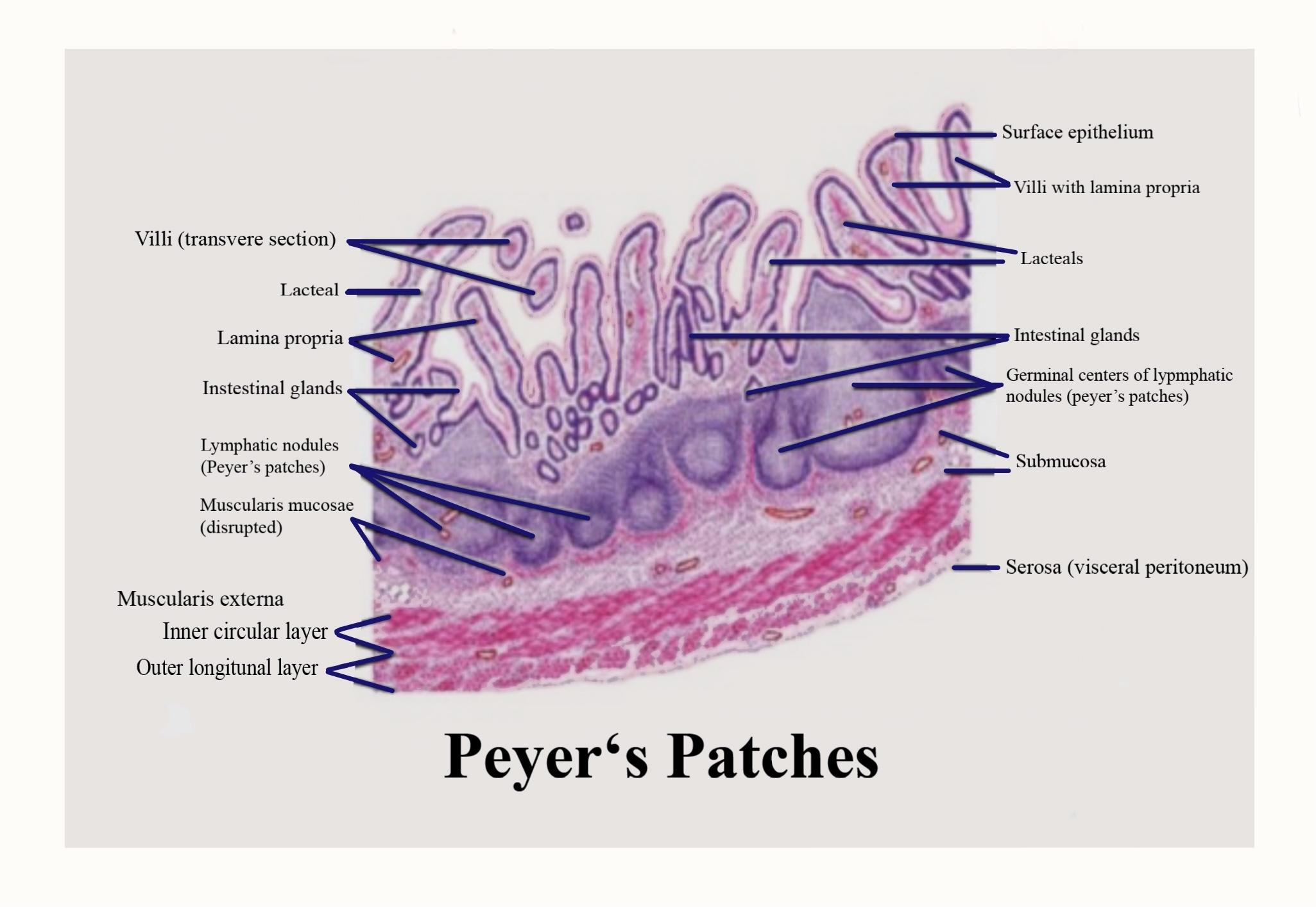
Peyer's patches are not passive filters. They are active inductive sites for mucosal immune responses.
Unlike standard intestinal villi covered with digestive enterocytes and goblet cells that produce thick protective mucus, the dome epithelium covering a Peyer's patch, known as the follicle-associated epithelium (FAE), is specialized. It contains very few goblet cells. Mucus here is exceptionally thin. This strategic thinning lets luminal contents touch the surface directly.
Within the FAE sit microfold cells (M cells). These epithelial cells lack conventional microvilli and boast a deeply invaginated basolateral membrane that forms a "pocket" containing dendritic cells, T cells, and B lymphocytes.
M cells sample antigens from the gut lumen via transcytosis. Intact proteins, dead commensal fragments, and live pathogens pass directly through the M cell body and are delivered directly into the hands of awaiting dendritic cells in the subepithelial dome. From there, antigen-presenting cells move into adjacent lymphoid follicles, priming naive T cells and stimulating B cells to undergo class-switch recombination into protective dimeric immunoglobulin A (dIgA).
Through this targeted antigen sampling, the host continuously profiles gut contents without triggering destructive, full-scale systemic inflammation.
David Miller brings 15 years of experience in global economics, personal finance strategy, and market dynamics. He specializes in turning complex economic trends into actionable insights for everyday readers.
